Indian Scientists Create Detailed Human Brainstem Atlas
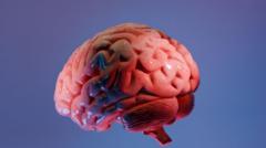
Scientists at the Sudha Gopalakrishnan Brain Centre (SGBC) at the Indian Institute of Technology, Madras, have developed Anchor, a highly detailed three-dimensional atlas of the human brainstem at cellular resolution. The atlas combines over 500 tissue sections from brains of individuals at various life stages, identifying more than 200 clusters of brain cells and nerve pathways using eight chemical markers.
This digital map bridges the gap between broad medical imaging and detailed cellular pathology, allowing researchers to seamlessly move from MRI scans to individual nerve cells. The SGBC team built Anchor using high-resolution microscope images, making the detailed mapping process more affordable than molecular techniques.
The freely available online atlas aims to become a key resource for neuroscientists, neurologists, and neurosurgeons, potentially aiding in understanding and treating neurological disorders like Alzheimer's, Parkinson’s, and stroke. Researchers plan to expand the atlas by imaging over 100 more brains to study disease progression at a cellular level.
Surfaced by the Discovery lens — one of the vital signs ovr.news reads.
How we evaluated this
AI summary
read the original for the full story — Read on bbc.co.uk . How we work →